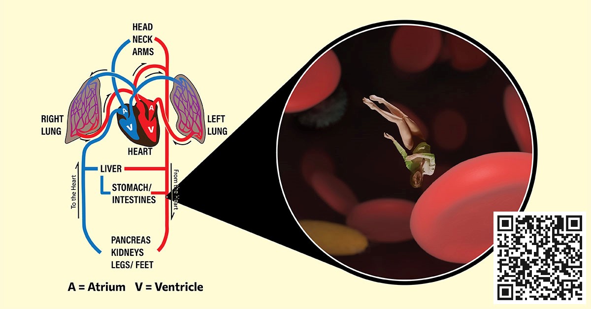
The circulatory system describes the heart and blood vessels, as they distribute blood to every part of the body.
Another name for this “heart (cardio-)” plus “blood-vessel (vascular)” system is “cardiovascular” system.
This article is in two parts:
- First, a poem in the form of a narrative of a person trapped within the circulatory system (press play below, or read the transcript that follows).
- Second, an explanation/ breakdown of the poem.
The Circulatory System (Red Prison)
Somehow, in a strange pool I got submerged;
not drowning, but panicky as adrenaline surged
It felt like an entrapment in a flooded subway,
with strong currents taking a one-way,
towards a snapping door, like a whale’s mouth
No escaping, and no telling north from south
As eyes gradually adjusted to situation,
to environment, I began to pay attention
Multiple creatures in a sea of amber,
with holeless red donuts having the number,
casting their color to surrounding yellow,
and in the distance, rhythmic bellow
In a swoosh, we were swallowed, like Jonah
powerless to swim like victims of belladonna,
dumped in a chamber, busy like a city hub,
its walls moving, yelling lub-dub, lub-dub
and urging us towards a three-leafed door
Resignedly, I accepted whatever lay in store
Into another chamber, we were coerced,
this one louder and scarier than the first;
its rough, expanding walls
with embedded crypts like shanty stalls,
closing in forcefully, as with malicious intent,
to extrude or maybe crush all content
Again discarded like cheap castaway,
into another tunnelly passageway
leading from the deafening lub-dub place
to a spirited path that split into a maze,
making the donuts glow like huge fireflies;
but alas, back to lub-dub house, in reprise
Back to more or less same process;
from “city hub”, to “shanty-walled” recess
As usual, from former, the latter receives,
but bright red fluid, via door with two leaves
then expelled, to begin a very long journey
via pulsating tunnels, like a swimming tourney
Eons later, it’s obvious this is prison,
wading through waters of shades of crimson;
a repeating cycle of lub-dubs and catacomb,
with donuts and crooked pacmans on the roam;
A circulatory system of sort;
an unending sea without port
TO BE CONTINUED
The "Red Prison", explained
Background
Within the ‘circulatory system’ of “heart + blood vessels + blood”, blood carries nutrients from food, as well as oxygen from breathing, to distribute to the whole body.
Like a pumping machine in a city water supply system, the heart plays a central role in the circulatory system; see The water tower analogy of the cardiovascular(circulatory) system.
The blood vessels are of different types and sizes:
- Arteries (singular = artery) are blood vessels that take blood away from the heart to rest of the body. Bigger arteries branch off to become smaller ‘arterioles’.
- Veins are blood vessels that take blood to the heart. Smaller veins (the venules) merge together to form bigger veins.
- Capillaries are the smallest blood vessels in the body, linking arterioles to venules. They are everywhere nutrients and gas exchange take place, because they have thin walls that permit these exchanges.
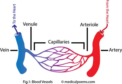
The circulatory system has two divisions, which are, the pulmonary circulation and the systemic circulation. The pulmonary circulation forms a circuit between the heart and the lungs, while the systemic circulation forms a circuit between the heart and the rest of the body.
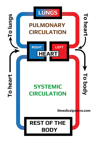
Learn about the heart here: THE HEART THAT BEATS (FOCUS ON THE NORMAL); I have also included #TheHeartPoem.
Stanzas 1 and 2 (inside the circulatory system, on the way to the heart)
“Somehow, in a strange pool I got submerged; … not drowning, but panicky as adrenaline surged … It felt like like an entrapment in a flooded subway, … with strong currents taking a one-way, … towards a snapping door, like a whale’s mouth … No escaping, and no telling north from south”
“As eyes gradually adjusted to situation, … to environment I began to pay attention … Multiple creatures in a sea of amber, … with holeless red donuts having the number, … casting their color to surrounding yellow, … and in the distance, rhythmic bellow”
Our fictional character, somehow is ‘inside’ the circulatory system, that is, inside one of the veins leading to the heart, likely the Inferior Vena Cava (IVC) (see below).
Blood is a “pool” of various components, including:
- Cells: Hole-less Donut(doughnut)-shaped Red Blood Cells (RBCs/ Erythrocytes), irregular White Blood Cells (WBCs/ Leukocytes) and discoid Platelets (Thrombocytes).
- Plasma: The liquid that holds all the components of blood, which is 90% water, plus other constituents, like glucose, proteins, hormones, carbon dioxide, and so on. The color of plasma without the cells, is amber/ yellow but the presence of Red Blood Cells, makes it appear red.
Every cell in the body depends directly, or indirectly on blood, to survive and function, because blood transports nutrients, like glucose, as well as oxygen to all the cells for use. It also helps to discard their waste products, like urea and carbon dioxide.
As the veins carry blood to the heart, the smaller veins keep merging together to form bigger ones; therefore, the biggest veins empty directly into the heart.
The Superior Vena Cava (SVC) and Inferior Vena Cava are the largest veins in the body, and they empty directly into the heart. The SVC carries blood from the head and arms, while the IVC carries from the abdomen and legs.
The IVC contains a one-way valve that prevents backflow/regurgitation of blood to the lower part of the body, which would otherwise happen because of gravity. The opening and closing of the IVC valve, could well be likened to a whale’s mouth, that is, if you are small enough to fit into the circulatory system.
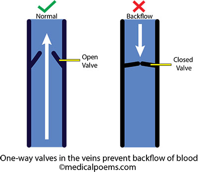
The SVC on the other hand, does not require this valve system, therefore it is “valveless”.
Other veins empty into the right atrium also, from parts of the heart muscles and lung tissue.
Stanza 3 (inside the right atrium)
“In a swoosh, we were swallowed like Jonah, … powerless to swim like victims of belladonna, … dumped in a chamber, busy like a city hub, … its walls moving, yelling lub-dub, lub-dub … and urging us towards a three-leafed door … Resignedly, I accepted whatever lay in store”
Blood from the SVC, IVC as well as veins that drain parts of the heart muscle and lung tissue, empty into the right atrium.
This blood is low in oxygen, however rich in nutrients, like glucose. It makes sense:
- Nutrients from the food we eat, enter into the circulatory system through the veins that drain the stomach (splenic vein) and intestines (mesenteric veins). These veins empty into the portal vein, which passes through the liver, then into the inferior vena cava (IVC), and into the right atrium.
- Oxygen enters into the circulatory system from the lungs (see below).
The structure of the right atrium is such that it is able to contract and expand, to expel it’s content into the right ventricle.
Atrial contraction is also known as “atrial kick”.
A one-way valve, known as the tricuspid valve (because it has 3 leaflets) lies between the right atrium and ventricle, and prevents backflow of blood from the ventricle to the atrium.
Stanza 4 (inside the right ventricle)
“Into another chamber, we were coerced, … this one louder and scarier than the first; … Its rough, expanding walls … with embedded crypts like shanty stalls, … closing in forcefully, as with malicious intent, … to extrude or maybe crush all content”
The right ventricle receives oxygen-poor blood from the right atrium, through the tricuspid valve. It has thicker walls than the right atrium, to help power its contractions, though its walls are thinner than the left ventricle’s.
It’s thick muscular walls enable it to expand and fill up with blood, as well as contract to expel blood through the ‘pulmonary trunk‘, to the lungs.
The inner lining of the right ventricle has irregular muscular ridges, known as trabeculae carneae.
Contractions of the right ventricle, ejects oxygen-poor blood to the lungs, through the pulmonary artery, for oxygenation.
Stanza 5 (through the pulmonary trunk, to the lungs)
“Again discarded like cheap castaway, … into another tunnelly passageway … leading from the deafening lub-dub place … to a spirited path that split into a maze, … making the donuts glow like huge fireflies; … but alas, back to lub-dub house, in reprise”
The pulmonary circulation is the part of the circulatory system that carries oxygen-poor blood from the heart to the lungs for oxygenation, and then back to the heart for distribution to the rest of the body.
When the right ventricle contracts, it ejects oxygen-poor blood through the ‘main pulmonary artery‘, also known as the ‘pulmonary trunk‘.
At the junction of the right ventricle and pulmonary trunk, is a one-way valve, ‘the pulmonic (or pulmonary) valve‘, which prevents backflow of blood to the right ventricle.
The pulmonary trunk divides into the right and left pulmonary arteries, which go to the right and left lungs, respectively.
Pulmonary arteries progressively divide into smaller ‘pulmonary arterioles’, and finally ‘pulmonary capillaries‘.
Air that we breathe in through the nose or mouth enters into the trachea (wind pipe), then into progressively smaller pipes (bronchi and bronchioles), all within the lungs. At the end of the smallest pipes are the alveoli, that look like bunches of grapes on the outside, but in actual sense, act like multi-lobed balloons.

Like balloons, alveoli inflate and deflate, as we breathe in and out; they can also hyperinflate and collapse, as seen in bullous emphysema, and atelectasis respectively.
The pulmonary capillaries more or less wrap around the alveoli, to maximize surface area for gas exchange.
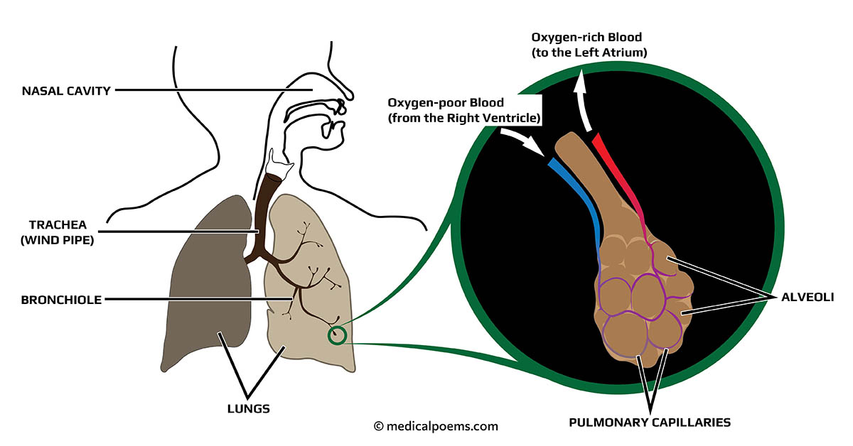
Gas exchange takes place across the thin layers of the alveoli and pulmonary capillaries. Oxygen molecules enter the blood, while carbondioxide molecules enter the alveolar space to be expelled, as we breathe out.
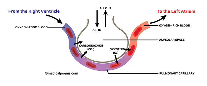
As the Red Blood Cells (RBCs) pass through the pulmonary capillaries, they take up oxygen; hemoglobin within the red cells bind to the oxygen molecules. Oxygen plus hemoglobin becomes oxyhemoglobin, which is bright red.
NOTE: Bright red colored RBCs carrying oxyhemoglobin gets distributed to the whole body through the arteries. As they get to the capillaries within the various organs however, the oxygen separates from the hemoglobin. Hemoglobin without attached oxygen is known as deoxyhemoglobin (deoxygenated hemoglobin); blood in the veins is dark red because of deoxyhemoglobin.
Oxygen-rich blood exits the lungs through the Pulmonary veins, thence to the left atrium of the heart.
In illustrations, we generally represent veins as blue, and arteries as red.
Stanza 6 (the left heart)
“Back to more or less same process; … from “city hub” to “shanty-walled” recess, … As usual, from former, the latter receives, … but bright red fluid via door with two leaves, … then expelled, to begin a very long journey … via pulsating tunnels, like a swimming tourney”
The “Left Heart” (Left Atrium plus Left Ventricle) functions much like the “Right Heart” (Right Atrium plus Right Ventricle). The major differences though, between the ‘left’ and ‘right’ heart, are:
- the left atrium receives oxygen-rich blood from the pulmonary veins. The right atrium on the other hand, receives oxygen-poor blood from the whole body.
- between the left atrium and left ventricle is the bicuspid valve, which has two leaflets. Conversely, between the right atrium and right ventricle is the tricuspid valve, which has three leaflets.
- the left ventricle is more muscular than the right, because it needs to generate enough force to distribute blood round the body.
Oxygen-rich blood is bright red, unlike the dark red oxygen-poor blood (see above).
Two pulmonary veins exit each lung (total = 4), but they merge to form two, which then empty into the left atrium.
From the left atrium, blood enters into the left ventricle. The powerful contractions of the left ventricle, forces blood through the aortic valve, into the ascending aorta (a large artery).
The cyclical filling up, and ejection of blood means that the heart acts like an intermittent pump, which takes in, and ejects blood in “batches”; however, it occurs so rapidly that blood flow is continuous and is apparent only as artery pulsations.
The right and left ventricles contract at approximately the same time, similar to the right and left atria (singular: atrium).
Stanza 7 (reality of the Red Prison)
“Eons later, it’s obvious this is prison, … wading through waters of shades of crimson, … a repeating cycle of lub-dubs and catacomb, … with donuts and crooked pacmans on the roam; … a circulatory system of sort, … an unending sea without port”
Practically every organ in the body receives oxygen-rich blood, through the same basic structure:

Once the organs use up the oxygen and nutrients, they empty their waste into the capillaries, for recycling thus:

This means that the circulatory system is a ‘closed system, not unlike a “prison”.
This cycle continues, for as long as the heart continues to beat.
NOTE: White Blood Cells and Platelets are also important components of the circulatory system.
- The White Blood Cells help to fight infections, for example by engulfing bacteria (like pacman).
- Platelets help to seal up breaks in the blood vessels, for example, during injury that cause bleeding.
Conclusion
The circulatory system is a network of heart + blood vessels, through which blood flows. It has two divisions, which are the pulmonary circulation and the systemic circulation.
Arteries, veins and capillaries make up the blood vessels.
The arteries take oxygen-rich blood from the heart to the organs and veins return oxygen-poor blood to the heart. Capillaries, with their thin walls allow gas exchange within the organs, and interconnects arteries and veins.
Oxygen-poor blood that enters the ‘right heart’ is sent to the lungs to make it oxygen-rich. Oxygen-rich blood then returns to the ‘left heart’, from where it gets distributed to the whole body.
The circulatory system is a closed system, also known as “the red prison”.
