The heart is an organ in the chest (thoracic) cavity, that helps to circulate blood, through its pumping action. It is a major component of ‘the circulatory system‘, which also includes the blood vessels (arteries, veins and capillaries), and blood.
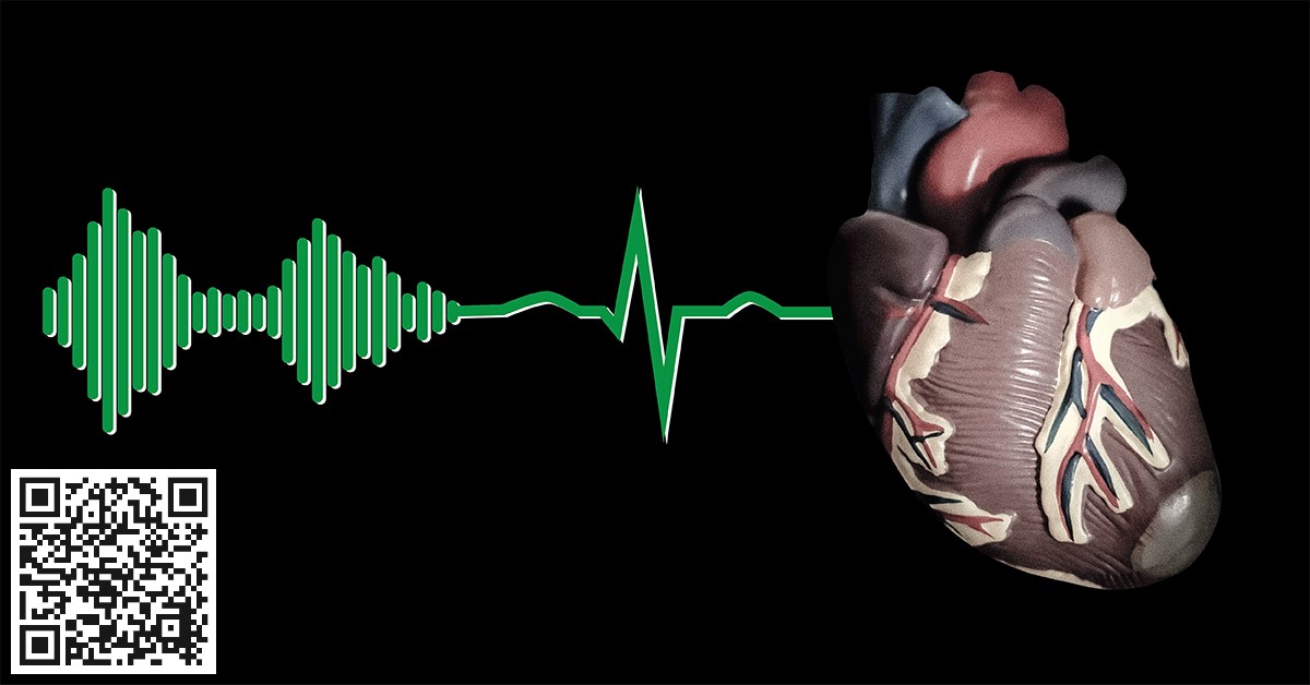
Below is a poem about normal functioning of the heart; I call it “the heart poem”.
Enjoy this classic poetry (video and transcript), and read this article to the end, to learn about the heart’s structure and function.
The Heart Poem
The heart, the size of a closed fist,
a vital organ, because it ensures you exist
Lub-dub, lub-dub it pumps away,
every second, everyday
Its layered walls enclose four cubicles,
the right and left atria and ventricles
Lub-dub, lub-dub it contracts and relaxes,
distributing through blood, nutrients and gases
Whether you’re five, or ninety-five,
it keeps the body healthy and alive
Lub-dub, lub-dub it’s reassuring beat,
as it pumps blood to head and feet
Some say it gives love, an emotion to treasure;
I say it pumps blood, maintaining its pressure
Lub-dub, lub-dub it regulates flow,
not too high, not too low
At work or asleep, it keeps pumping;
with stress or distress, it starts thumping
Lub-dub, lub-dub becomes faster
If unsure, call the doctor
To keep the heart properly functional,
a healthy diet and exercise are not optional
Lub-dub, lub-dub the heart should sound,
like a talking drum on a merry-go-round
The Heart Poem, explained
Stanza 1 (Introduction: The heart as a pump)
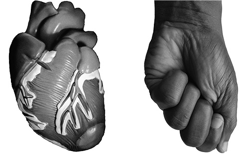
“The heart, the size of a closed fist,” (stanza 1, line 1)
The heart is an organ in the chest cavity, that lies between the right and left lungs.
Most texts describe the heart as the size of a fist; this is true. It’s shape could also appear like that of a fist, if you make it like in the image below.
Research shows that males have slightly larger hearts than females, but this does not become apparent until puberty (follow this link to learn more).
“a vital organ, because it ensures you exist” (stanza 1, line 2)
Some organs in the body are called “vital organs”, because they are essential for survival. These organs include the brain, lungs, liver, kidneys and heart.
One can survive without an arm, spleen or reproductive organs, but not for long, without the vital organs.
“Lub-dub, lub-dub it pumps away, … every second, everyday” (stanza 1, lines 3 & 4)
Without the pumping action of the heart, blood cannot flow within the blood vessels (arteries and veins) (see The Circulatory System: A look into the Red Prison).
Let us see how this ‘pumping action’ of the heart comes about.
- A little structure known as the sinoatrial (or SA) node, lies within the walls of the heart. This ‘SA node’ acts like a little brain by initiating and sending electrical signals; hence it is known as the pacemaker. Asides being able to initiate and send out signals on its own, it also responds to signals from the brain.
- The electrical signals cause the muscles of the heart to contract and therefore squeeze out blood; see details below.
- Before the next signals come from the SA node, the heart fills up with blood again, and the cycle continues.
- At rest, the SA node “fires” the electrical signals about 60-100 times a minute, that means at least one contraction every minute.
- Every time the heart contracts and forces out blood, the arteries expand or “pulsate“, in unison, to accommodate the inflow. “Pulsations” or “pulse” represent the heart rate; see below.
Anything that increases the heart rate, like exercise, fever, high caffeine consumption, etc, will make the pulse to be rapid.
Stanza 2 (Heart Anatomy)
“Its layered walls, enclose four cubicles, … the right and left atria and ventricles … “Lub-dub, lub-dub it contracts and relaxes, … distributing through blood, nutrients and gases” (stanza 2, lines 1 & 2)
The heart is like a house that has four rooms; one room leads to the next, and movement is in one direction only.
The rooms or “chambers” are the right atrium, right ventricle, left atrium and left ventricle.
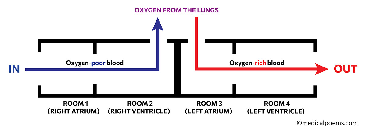
Though some parts are thicker than others, the general structure of the walls of the heart is:
- An outer layer, the epicardium
- A middle muscular layer, the myocardium: the type of muscle here, cardiac muscle is unique to the heart. Myocardium in the ventricles is thicker than in the atria.
- An innermost layer, the endocardium
- The pericardium is a separate resilient layer, that encloses the whole heart.
Chamber 1: The Right Atrium
Blood enters the right atrium through the largest veins in the body, that is the Superior Vena Cava (SVC) and Inferior Vena Cava (IVC).
The SVC carries blood from the head, neck and arms, while the IVC carries from the abdomen and legs.
Other veins also empty into the right atrium from parts of the lungs not involved in gas exchange (lung parenchyma), as well as heart muscles.
Blood entering the right atrium has low oxygen, because the organs have used up the oxygen for normal functioning.
The right atrium is able to expand to accommodate blood, and eject the blood into the right ventricle.
Although the walls of the right atrium are relatively thin, it contains parallel muscle fibers like the teeth of a comb (pectinate muscles), that help with contraction.
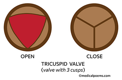
The right atrium ejects its content into the right ventricle through a ‘one-way’ (prevents back-flow) tricuspid valve.
Chamber 2: The Right Ventricle
‘Oxygen-poor’ blood from the right atrium enters into the right ventricle through the tricuspid valve, which has three flaps, that form a “Y”, where all three meet.
Irregular ridges line the right ventricle, unlike the right atrium that has mostly-smooth lining. These ridges, known as trabeculae carnae (meaty ridges) help with contractions directly and indirectly.
- Firstly, by supporting the underlying compact myocardium layer in contracting the ventricle.
- Secondly, by forming a band, the moderator band, which serves as a conduit for passage of nerve fibers that carry electrical signals; this also leads to contraction.
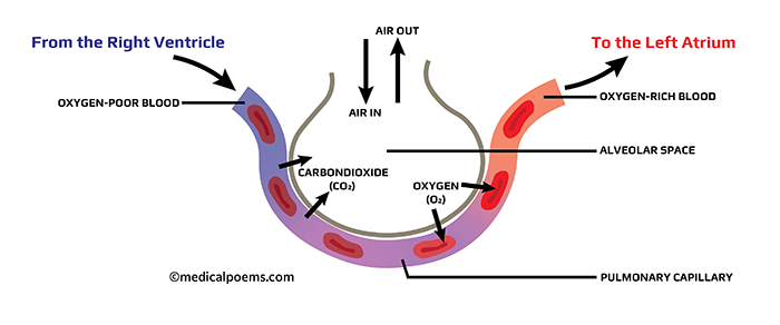
The right ventricle empties its content of oxygen-poor blood into pulmonary trunk/artery, which leads to the lungs, for ‘oxygenation’. Oxygenation is the addition of oxygen to the blood (See The Circulatory System: A look into the Red Prison)
Between the right ventricle and pulmonary trunk is the pulmonic (or pulmonary) valve, which has three flaps, each half-moon-shaped.
Chambers 3 & 4: The Left Atrium and Left Ventricle
The left atrium and left ventricle make up “the left heart“, as against “the right heart“, which is the right atrium plus right ventricle.
Oxygen-rich (oxygenated) blood from the lungs, returns to the left atrium, then to the left ventricle.
Structurally, the left atrium is similar to the right atrium; also, the left ventricle is similar to the right ventricle.
Between the left atrium and left ventricle however, is a valve that has two cusps, therefore known as the bicuspid valve.
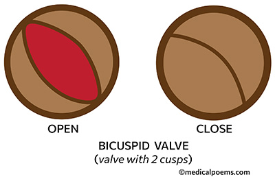
The left atrium has fewer pectinate muscles, while the left ventricle is more muscular than the right, because it needs to generate enough force to distribute blood round the body.
When the left ventricle contracts, it pumps out oxygen-rich blood through the aorta, the largest artery in the body.
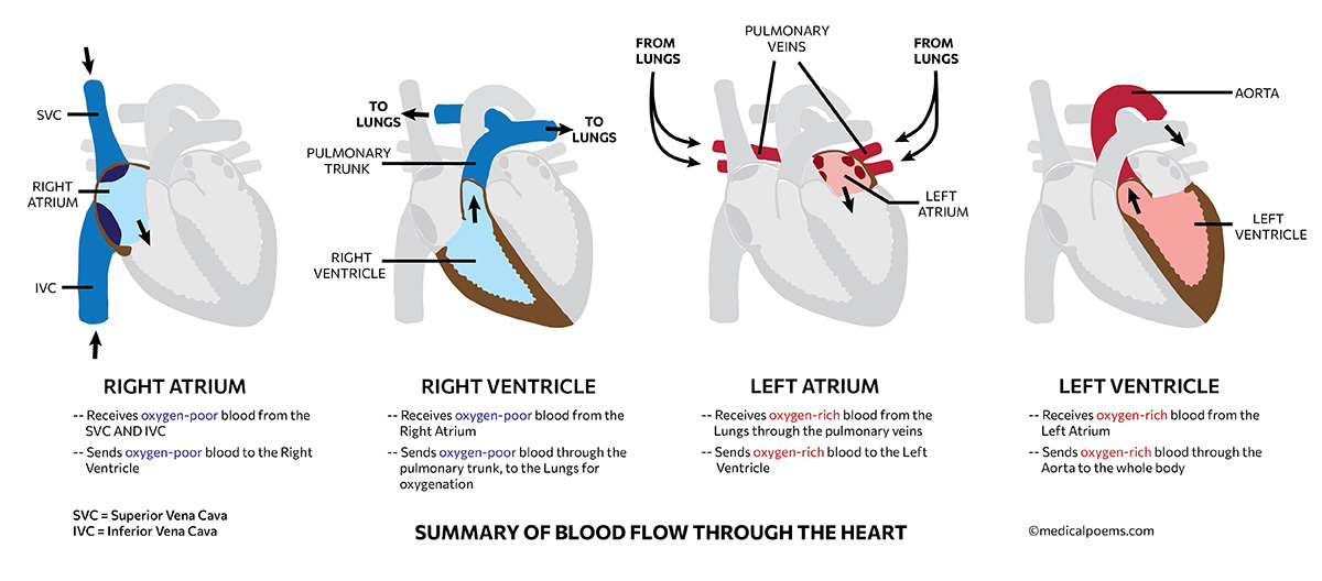
Between the left ventricle and aorta is an “aortic valve“, which has three flaps, each half-moon-shaped, like the pulmonic valve.
Stanza 3
“Whether you’re five, or ninety-five, … it keeps the body healthy and alive … Lub-dub, Lub-dub its reassuring beat, … as it pumps blood to head and feet” (stanza 3, lines 1 to 4)
The heart is the first organ to become functional, in the developing fetus which is as early as five weeks after the mother last sees her menses and it continues to beat throughout life.
Many advanced countries have criteria to define death, which includes, the heart stopping to beat, respiration ceasing, and/or brain death. In most cases however, once there is irreversible cessation of breathing or normal heart activity, death is diagnosed.
Stanza 4 (Blood Pressure)
“Some say it gives love, an emotion to treasure; … I say it pumps blood, maintaining its pressure … Lub-dub, lub-dub it regulates flow, … not too high, not too low” (stanza 4, lines 1 to 4)
Normal heart activity involves repeated cycles of ‘contraction’, to squeeze out blood through the arteries, as well as ‘relaxation’, to take in blood from the veins.
Systole is the state of the heart when it contracts, while Diastole is it’s state when it relaxes.
At systole, the force of contraction of the heart creates maximum pressure in the arteries; therefore, this pressure is known as systolic blood pressure. On the other hand, the pressure of blood in the arteries when the heart is relaxed at diastole is the diastolic blood pressure.
Blood pressure is a measure of systolic and diastolic pressure, written as:
An example of blood pressure reading is:
The body tries to maintain blood pressure within a particular level because high blood pressure (hypertension) or low blood pressure (hypotension) has detrimental effects on the body.
Read about hypertension here: Hypertension: focus on the tactics of the silent killer.
While the heart itself does not directly regulate blood pressure, it responds to feedback from the brain, which regulates the speed and force of contraction, as well as vascular resistance.
The kidneys and adrenal glands also play major roles in regulating blood pressure.
Stanza 5 (Heart sound, rate and disease)
“At work or asleep, it keeps pumping; … with stress or distress, it starts thumping … Lub-dub, lub-dub becomes faster; … if unsure, call the doctor” (stanza 5, lines 1 to 4)
As the healthy heart works, it makes two audible sounds, S1 and S2, which we colloquially call Lub-Dub (S1=Lub; S2=Dub).
The first heart sound, S1 is from the tricuspid and bicuspid valves closing when the ventricles contract in systole. S2 (second heart sound) on the other hand, is from the closure of the pulmonic and aortic valves when the ventricles relax in diastole.
Note that the tricuspid and bicuspid valves are known as the atrioventricular valves because they interpose the aria and ventricles; tricuspid on the right, and bicuspid on the left (see above).
The pulmonic and aortic valves on the other hand, are known as the semilunar valves because the valve flaps have a half-moon shape.
Additional sounds, S3 and S4, may be present in disease states, like when the valves do not function properly.
Heart rate is the number of times the heart beats in a minute, that is the number “S1-S2 (Lub-dubs)” in a minute; the normal is 60-100 (average 72) beats per minute (bpm). Bradycardia is a heart rate less than 60bpm, while tachycardia is greater than 100bpm.
Conditions like stroke, hypothyroidism and cardiomyopathy may cause bradycardia, while exercise, use of stimulants and anxiety, may cause tachycardia.
An easy way to measure the heart rate is by checking the pulse; place the pads of the middle and index fingers at the wrist, just under the thumb and press lightly. You should feel a rhythmic push, as the ‘radial artery‘ underneath, “pulsates“.
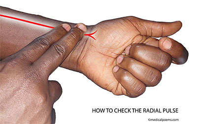
Get a clock or stopwatch and count the number of ‘pulses’ in one minute; that is the heart rate.
Generally, it is not obvious, that your heart is beating, but in certain conditions, you may become aware that your heart is beating harder, faster (tachycardia) and/or irregularly; these are palpitations.
Palpitations may come with obvious chest thumps or fluttering, rapid/bounding pulse, or pounding sensation in the chest or neck. It may be due to anxiety, panic disorders, drug abuse, thyroid dysfunction, side effects of medications and so on.
Stanza 6 (Effect of diet and exercise on the heart)
“To keep the heart properly functional, … a healthy diet and exercise are not optional … Lub-dub, lub-dub the heart should sound … like a talking drum on a merry-go-round” (stanza 6, lines 1 to 4)
Research shows that a healthy diet and exercise are important for mood, good sleep, memory, as well as bone and muscle health.
Most importantly however, they are important for prevention or reduction of severity of chronic illnesses, like hypertension, diabetes and heart disease. These conditions have direct and/or indirect bearing on the heart.
Conclusion
The heart is a vital organ in the chest cavity, that helps to distribute oxygen and nutrients through blood.
It has a robust muscular layer, that helps to power its contractions, as blood moves through four chambers, namely left and right ‘atrium’ and ‘ventricle’.
The heart also has atrioventricular and semilunar valves that help to prevent backflow/ regurgitation of blood.
We may be prone to conditions like hypertension, diabetes and heart diseases, if we do not take healthy diet or exercise regularly.
